We all know that sensory cortices have “maps” of the sensory world, such as the famous homunculus. But where do these maps come from? It was thought that these maps form when thalamic projections wire into the cortical input layer and refine from an initially very messy state into clean maps based on sensory experience. The cortical input layer is also called layer 4 and is the middle layer of the cortex. Now there is a wrinkle in development: at the earliest stages, thalamic fibers do not connect to layer 4 right away but connect to cells deep in the cortex called subplate neurons. In humans, primates, and cats, these cells are transient — most die off during development. The role of these subplate neurons during their existence has been enigmatic.
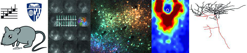
Travis created a genetically engineered mouse (Cplx3-CreER) that let us selectively image the activity of these neurons. Using calcium imaging to watch these neurons fire in living mouse pups, he and Minzi found that at very young ages, subplate neurons receive spontaneous activity from the peripheral sensory organs such as the eyes and ears. They further found that topographic protomaps were present even before the onset of sensory-driven experience.
Together, we think subplate neurons form a protomap of cortical organization by integrating thalamic inputs and then distributing them across developing cortical circuits. Thus, “maps” form earlier and differently than we thought, and we think that subplate neurons help establish or sculpt the connectivity patterns in the adult cortex that underlie normal function
This long-term, complex project would not have been possible without the lab’s fantastic teamwork!!!
Read it here in Nature Communications
